For more detailed information about your recovery, you can download the Take Heart: Your Heart Your Recovery booklet (PDF).
On this page
High Blood Pressure
What is blood pressure?
Blood pressure is the force that circulating blood puts on the artery walls. When blood pressure is high, there is more pressure on the artery wall than usual. Some people have high blood pressure and do not know they have it.
The extra pressure damages the smooth lining of the arteries and makes it easier for cholesterol and fat to build up along the artery walls. As the arteries become clogged with these fatty layers (atherosclerosis), less blood gets through. This causes the heart to beat harder as it tries to pump blood through narrowed arteries. If untreated, high blood pressure may in time damage the heart, brain and kidneys. It is a leading cause of heart attacks and strokes, heart or kidney failure. The exact cause of high blood pressure is not fully known for many people. High blood pressure can be treated and lowered with medication.
How do I control blood pressure?
There are a number of ways to reduce blood pressure, these include:
- Get your blood pressure checked as often as your doctor suggests
- Stop smoking
- Take your medications as regularly as prescribed
- Reduce your salt intake and try to avoid processed, convenience and fast foods. The easiest way to reduce salt intake is not to add salt at the table or in cooking
- Fruit and vegetables provide us with a good source of potassium which can help control your blood pressure. Aim for five portions a day
- Reduce stress by learning ways to relax and by exercising.
- Lose weight if needed. The heart pumps harder to supply an overweight body with blood and oxygen.
Cholesterol
Cholesterol and triglycerides are fatty chemicals in the blood. There are two main types of cholesterol; LDL ‘bad cholesterol’ which carries cholesterol from the liver to the rest of the body, and HDL ‘good’ cholesterol which returns excess cholesterol to the liver. If you have high levels of cholesterol and triglycerides, your risk of coronary heart disease is greater. High levels of LDL cholesterol stick to the walls of your arteries and make plaque. This plaque blocks the arteries, interfering with the blood flow which can make a heart attack more likely. Most people with high blood cholesterol will only need advice about diet, a healthy lifestyle and possibly blood cholesterol monitoring.
Angina
What is angina?
Angina is a discomfort or pain in or adjacent to the chest, which is due to a reduced supply of blood to the heart muscle.
What is the cause of angina?
The exact cause of angina is unknown. However, we do know that certain factors make some people more likely than others to get angina. These are called “risk factors”. The term “risk factor” is used to describe a feature that is associated with the development of coronary artery disease or coronary heart disease.
Coronary heart disease results from narrowing of the coronary arteries. Because of the narrowing, the heart muscle may not receive enough blood, which contains oxygen and nutrients. It is this lack of oxygen within the blood that causes pain.
In the majority of cases coronary heart disease is the result of a process called atheroma or atherosclerosis. Another name you may be familiar with is “furring up” or “hardening of the arteries”. What this means is fatty deposits called plaque accumulate in the coronary arteries causing narrowing. This process is slow and starts in young age.
The major “risk factors” for developing coronary heart disease are cigarette smoking, high blood cholesterol and high blood pressure. Other risk factors are obesity, stress, diabetes, lack of exercise and family history of heart disease.
It is important to recognise that there is no single cause of coronary heart disease but the more risk factors you have, the more likely you are to develop the disease.
What is the treatment of angina?
Treatment is divided into two parts.
Treatment started by you:
Identifying and modifying the risk factors where possible (family history is an unmodifiable risk factor).
In the long term this is the most important aspect of treatment and will involve giving up smoking, checking blood cholesterol, checking blood pressure and if necessary, lowering cholesterol and blood pressure. You can ask your G.P. to check these.
Treatment started by the doctor:
Medications prescribed for angina, angioplasty and heart bypass surgery.
Medications can help in two ways, they can either improve the blood flow through the coronary arteries or they can reduce the work the heart has to do. Some of the medications commonly used to treat angina are:
Aspirin
Reduces the stickiness of blood platelets and therefore the tendency for the blood to clot.
Beta-blockers
Reduce the work of the heart by regulating the heartbeat and slowing it down, as well as reducing blood pressure. Examples include atenolol, metoprolol, propanolol and sotalol.
Calcium antagonists
Help to prevent angina in two ways; they relax and dilate the coronary arteries therefore allowing more blood to reach the heart muscle and helping the heart muscle to use the oxygen and nutrients carried by the blood more efficiently. Examples include nifedipine, diltiazem and vesapamil.
Nitrates
Relax the smooth fibres of the blood vessels, thus allowing the coronary arteries (as well as other arteries in the body) to dilate. Examples include isosorbide mononitrate and isosorbide dinitrate.
There are two types: short acting and long acting.
Short acting: GTN spray or tablet put under the tongue for quick absorption, giving relief within a minute or two.
Heart Attack
Symptoms of a heart attack are similar to angina. A heart attack occurs when the blood supply to the heart muscle is blocked for a long period of time (due to one or more blocked coronary arteries). The blockage is often caused by a blood clot. This results in permanent damage to the heart muscle beyond the area of blockage. Symptoms of a heart attack usually last longer than 30 minutes and are not relieved by rest of medication, such as GTN spray. A heart attack may be the first sign of coronary heart disease.
Pacemaker
A pacemaker is a battery operated device, inserted into the body just below the collar bone. A wire or ‘electrode’ leads into the heart.
The most common pacemaker is designed to ‘sense’ the speed of your heart beat. If the rate falls below a certain level, the pacemaker ‘senses’ this and sends impulses along the electrode to stimulate or ‘pace’ the heart beat at a faster more appropriate rate until your own heart beat increases again.
There are many different types of pacemakers which are individually selected for your particular needs.
Why do I need a pacemaker?
There are many reasons why people may need a pacemaker. If your pulse falls to a slow rate you could feel dizzy, tired and sleepy. You may even have been experiencing blackouts which can lead to personal injury. Some people experience a fast erratic heart rate causing ‘palpitations’. You may also feel breathless. It is also possible not to experience any of the above but your doctor may still advise a pacemaker.
Please discuss your nurse / doctor which type of pacemaker you have and how it will help your symptoms.
Implantable Cardioverter Defibulator
ICD’s are implanted to protect from serious fast heart rhythm disturbances (arrhythmias). Serious or life threatening arrhythmias are usually from the lower chambers of the heart, there are two specific fast heart rhythms (Tachycardia’s) called VT (ventricular tachycardia) and VF (ventricular fibrillation). Arrhythmias can occur for many reasons, they are difficult to treat in that they can occur at random and it is often impossible to identify a specific triggering factor. In general they occur more commonly in patients who have had previous heart damage or structural changes to the heart muscle, though they can at times occur in patients whose heart muscle is structurally normal.
Treatment options are limited for these dangerous type of rhythm disturbance, medication is often used to prevent or reduce the occurrence of an arrhythmia, with the ICD there is a back up.
The ICD is a small device containing a battery and computer; it differs from an ordinary pacemaker because it has the ability to deliver large electric shocks. It is usually implanted in the left chest wall under the collarbone and connects to the heart via 1, 2 or 3 leads or wires. Its job is to constantly monitor the heart rate. Should it detect a fast rhythm it can deliver electrical therapy to “reset” the heart back into a normal rhythm.
Patients who are considered for this type of device have either experienced a serious arrhythmia or are likely to do so. Your nurse or doctor can explain how it applies to you.
ICD’s are mainly aimed at treating electrical problems in the heart, in general they will not alter other cardiac symptoms; for example chest pain or breathlessness.
What is the difference between an ICD and a pacemaker?
Pacemakers are much smaller than ICD’s but use similar technology and are often made by the same companies. They are used to treat slow heart rhythms (Bradycardia) this may occur due to Sinus Node Disease or when the heart is failing to adequately transfer the electrical impulse “Heart Block”. Pacemakers provide a small electrical impulse to stimulate the heart muscle to contract. They can be programmed in many ways to provide these impulses as and when required or pace all the time. They unfortunately do not protect against fast rhythm problems. ICD’s on the other hand can provide a pacemaker function in addition to being able to treat fast rhythm problems.
How does the defibrillator work?
ICD’s constantly monitor the rate and rhythm of the heart. The rate at which they are set to detect fast rhythms vary from one individual to another. They can be adjusted further at out patient appointments if the pattern or the speed of arrhythmia changes.
Should the ICD detect a fast heart rate it will either try to “Anti tachy pace” or “fast pace” the heart to try and interrupt the arrhythmia, this is often painless with most patients being unaware of it happening, if this is unsuccessful or if the heart rate is very fast it will deliver an electric shock to try and reset the heart. The shock lasts less than a second but it is painful and can make you jump. Patients with ICD’s often describe it as a “kick in the chest”. When patients have felt a shock it is common to feel a little frightened at first but this usually settles down very quickly. Rarely it may need to shock more multiple times.
Heart Failure
Heart Failure does NOT mean that the heart has or is about to stop working. Heart Failure occurs when the heart fails to pump out enough blood to meet the demands of the body. It can often result in the build-up of fluid in the lungs or other parts of the body, most commonly the legs or stomach. This is often associated with breathlessness.
What causes heart failure?
Many things can cause heart failure. The most common causes are:
- Heart attacks (causing damage to heart muscle)
- Coronary Artery Disease (Angina)
- Heart Valve Disease
- Disease of the heart muscle (Cardiomyopathy)
- Hypertension (high blood pressure)
- Infections of the heart
- Abnormal heart rhythms
- Alcohol or drug abuse
- Congenital heart disease (an abnormality of the heart at birth)
What is the treatment of heart failure?
In the majority of cases Heart Failure cannot be cured, it is a chronic condition. However, taking certain medication and making lifestyle changes can greatly improve the symptoms and slow the progression of Heart Failure. It can also help you to gain some control over your condition.
More information can be found in the Take Heart Heart Failure booklet (PDF).
Infective Endocarditis
Endocarditis is caused by an infection, which may affect one or more of the heart valves and the lining of the heart muscle known as the “endocardium”. It is uncommon, but not rare, affecting 5 out of every 100,000 people and is often very serious. It occurs when a large number of bacteria in the bloodstream are attracted to the heart valve and begin to multiply. The bacteria stick together to form a clump which is known as “vegetation” which becomes attached to the heart valve, damaging it and causing it to malfunction. Vegetation can also form on artificial materials located within the heart such as artificial heart valves, pacemaker leads, dialysis lines and Hickman lines. This infection of the heart valve can spread through the bloodstream to other parts of the body. When part of a vegetation becomes detached it may spread to the brain, lungs, kidneys, spine or elsewhere.
What causes it?
There are numerous ways that the bacteria can enter our bloodstream. They can enter our bloodstream from a variety of sources, including from the bowel, having a dental procedure, poor dental hygiene, minor cuts or surgical incisions or invasion of the blood vessels by needles. Endocarditis usually results from a build up of the bacteria which the body’s immune system fails to destroy appropriately. Usually the body’s defence mechanism will destroy bacteria. However, if there are sufficient numbers they can collect and accumulate on the heart valves. Infection rarely occurs on normal valves. Valves that have been damaged in some way or are not working correctly due to rheumatic fever, congenital heart disease or previous endocarditis are more prone to developing infections. Additionally artificial valves and valve surgery can increase the risk. Injection drug abuse is a common risk factor for developing the condition. However, for many patients there is no clear explanation for how the bacteria have entered the bloodstream.
More information about Endocarditis can be found in this booklet (PDF).
Cardiac MRI
The Leeds Cardiac Magnetic Resonance (CMR) service is based at the Yorkshire Heart Centre, at Leeds General Infirmary. The Unit was founded in 1999 with the installation of a British Heart Foundation funded MRI scanner. Since then, the facility has expanded to include three state-of-the-art MRI scanners in a Clinical Imaging Centre (Clarendon Wing) and an Advanced Imaging Centre (Gilbert Scott Building).

We are a multidisciplinary team of medical professionals and we scan and report approximately 2500 scans every year on patients with a wide range of heart diseases. We receive referrals from several secondary care hospitals in West, North and South Yorkshire and work closely with clinicians in Leeds and across the region.
What is a Cardiac MRI?
A cardiac MRI scan is a detailed scan of the heart using a technology called magnetic resonance imaging. The scan is very safe and has the benefit that it involves no radiation. It cannot be undertaken in people with some types of metallic implant or pacemaker devices.
Before your MRI
If you are having a scan, we will write to you with an appointment and any instructions for the day of the scan (e.g. sometimes the scan gives us better information if you avoid caffeine beforehand). You can otherwise eat and drink normally, and there are no driving restrictions.
You may be in the department for around two hours, including the scan itself. The scanner can feel a little close to your face and chest, but most people tolerate this very well and we will do our best to make it easier.
It would be very helpful if you could let us know in advance if you have any metallic devices, implants or any metal in your eye(s). Our contact details are below.
On the day of your MRI
A radiographer or assistant will take you through a safety questionnaire to ensure it is safe to go ahead and that any detachable metal is stored securely in a locker before we take you to the scanning room. For most scans, we need to give you a contrast dye which gives us clearer pictures of the heart. For some scans we also need to give you a medication that simulates the effects of exercise on the heart for a short time. For these we will insert a cannula into a vein in your arm. The radiographer will check your height and weight so the measurements of your heart are corrected for your body size.
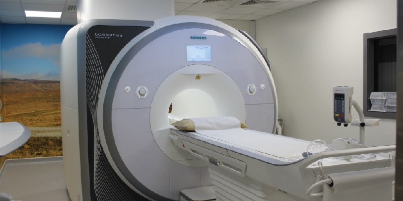
Once you are in the scanner room, we will place some soft stickers on your chest to measure your heart rate and rhythm and set you up for the scan. The machine can be noisy, so you will be given ear defenders and a buzzer and we can set up a radio station of your choice to distract you from the noise. You can see the scanner in the photo below; we will leave the room during the scan but can speak to you and hear you from the control room at all times.
Most heart scans require some breath-holding, so we will ask you when to breathe in, breathe out and hold, and when to breathe freely. Typically, a breath-hold lasts 5-10 seconds. Most cardiac MRI scans take between 30-60 minutes to complete and we will communicate with you throughout.
Once the scan is complete and the radiographer and/or doctor are happy with the images of your heart, you will be free to pick up your belongings and go. Our doctors will then report the scan and pass the report to the consultant who has requested the scan (usually your cardiologist).
Meet the team
How to contact us
- Leeds Teaching Hospitals NHS Trust: 0113 243 2799
- Cardiac MRI Bookings: 0113 392 5061